SEM-EDX Laboratory - Spherical Image - RICOH THETA

A material consists of phases, and a rock does mainly of minerals. To understand what the rock is, firstly we describe distribution and elemental abundances in minerals. Size of a mineral varies from meter- to nano-scale, and our nominal microbeam technique allows description of elemental distribution down to sub-micron scale.
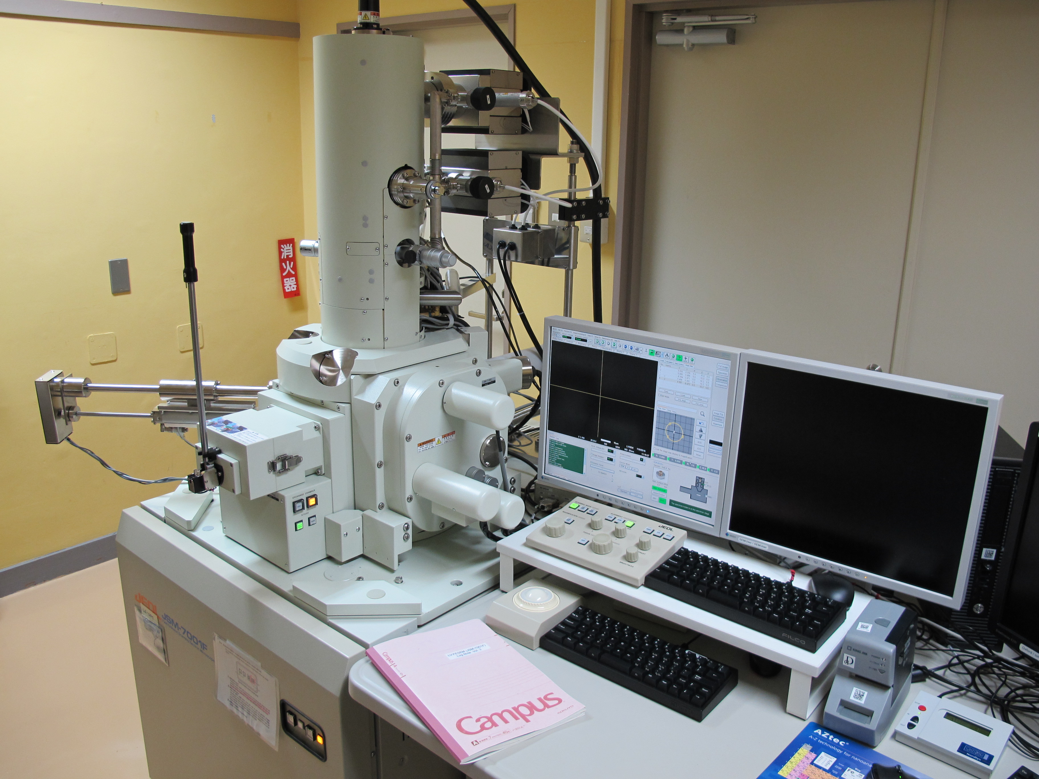
Following a nominal optical observation, we describe distribution of minerals using electron beam, and determine their major element compositions using X-ray spectroscopy, that is, scanning electron microscope (SEM) equipped with energy-dispersive X-ray spectroscope (EDS).
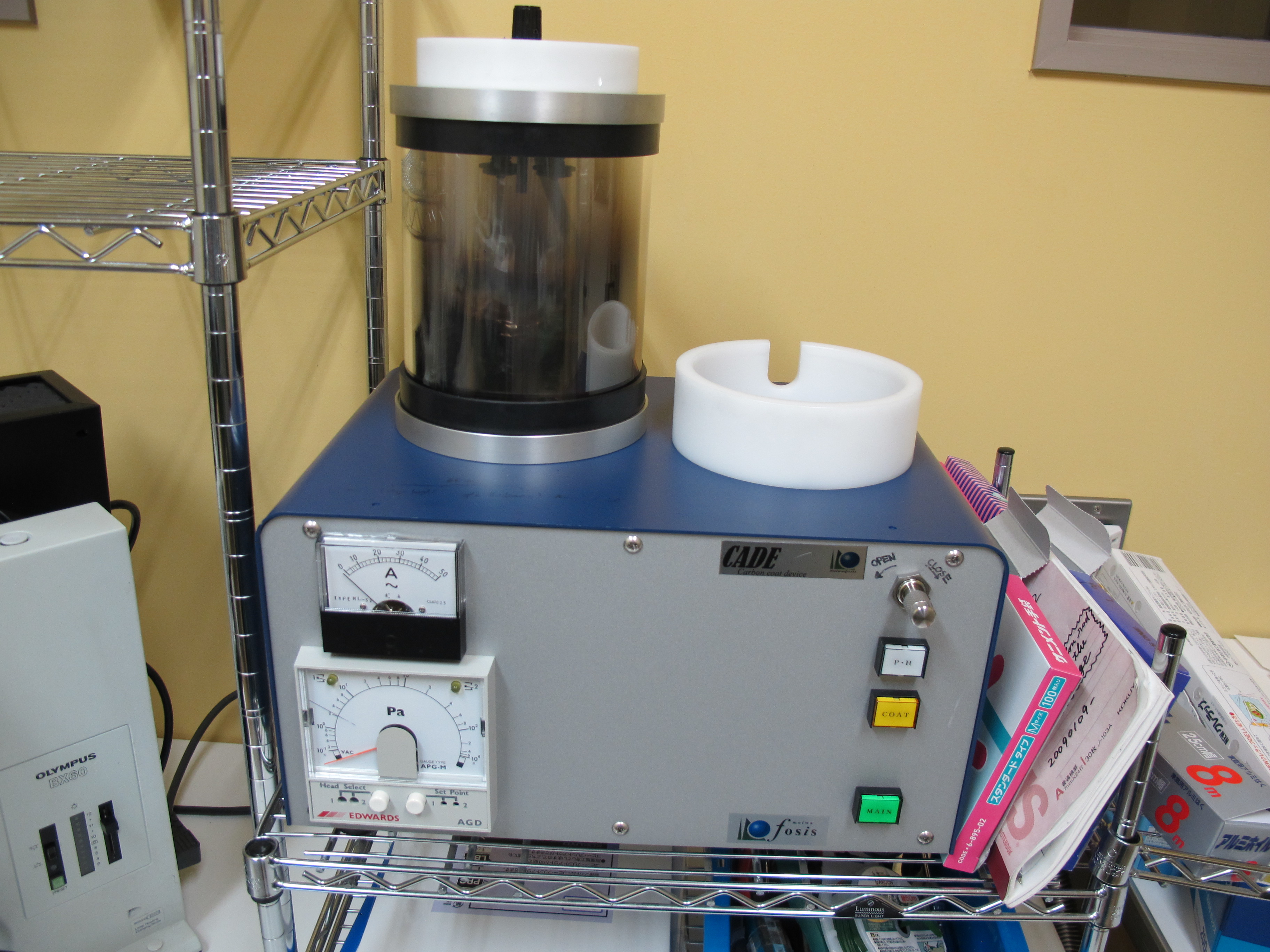
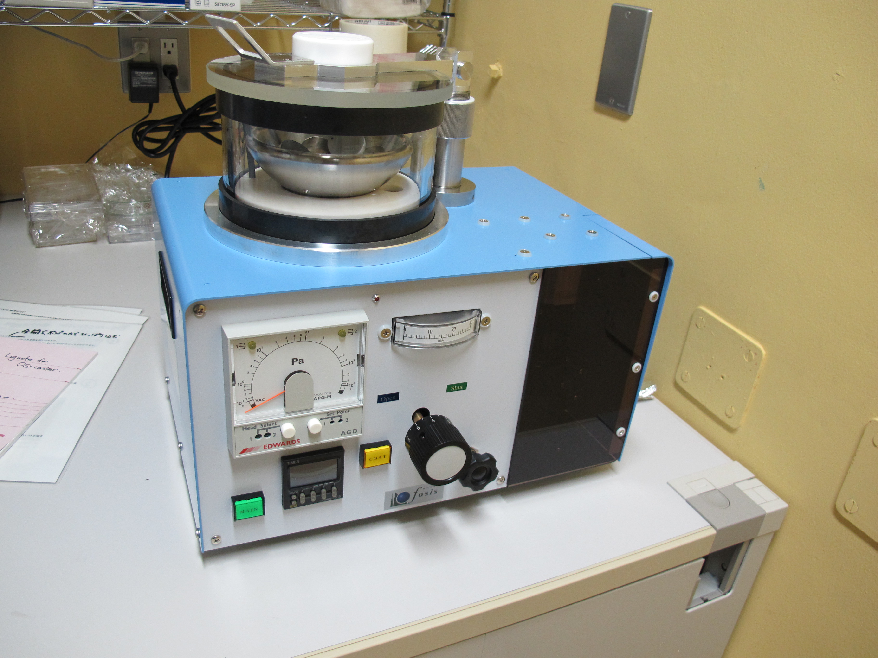
Since the probe analyses incident charged particles to the surface of the sample, coating to guarantee conductivity is necessary. We prefer carbon for electron probe, but if carbon-bearing materials are concerned, osmium is also available.
Our SEM has the field emission (FE) gun. The ordinary SEM has a thermionic emitter, which heats up a filament and emits electrons. The field emission is reached, not by heating, but by placing the filament in a huge electrical potential gradient, and can avoid the problems that the thermionic emitter involves; such as thermal drift during operation. The FE-SEM is a user-friendly instrument enable us to access highly stable and bright electron beam to achieve high spatial resolution images and elemental maps at submicron scale.